Уход после операции ревизии несвязанного эндопротеза плечевого сустава
а) Послеоперационное ведение и результаты:
- На протяжении ближайших двух суток после операции пациент остается в стационаре. На следующий день после операции начинается физиотерапия, однако ее характер зависит от типа выполненной ревизии:
• Пациентам, которым не выполнялась пликация задней капсулы, назначаются упражнения I фазы, направленные на растяжение сустава. Пределы движений зависят от особенностей операции. Мы рекомендуем своим пациентам пассивный подъем плеча в плоскости лопатки и пассивную наружную ротацию с использованием блоков или маятников. Активная внутренняя ротация исключается до шести недель после операции, однако допускается пассивная внутренняя ротации до груди. При соблюдении названных ограничений пациентам разрешают пользоваться конечностью в повседневной жизни
• При ревизиях по поводу нестабильности характер допустимых движений определяется нагрузкой на швы капсулы и сухожилий
• После пликации задней капсулы сустава выполняется иммобилизация «гангстерским» брейсом, а подъем плеча допускается только в плоскости брейса в нейтральном положении или положении легкой наружной ротации
• Вне зависимости от характера операции разрешаются пассивные движения в кисти и локтевом суставе
- Пациенты осматриваются амбулаторно через две недели после операции. Во время этого визита оценивается состояние раны и контролируется соблюдение пациентом данных ему рекомендаций. В следующий раз пациенты осматриваются через шесть недель. В этот период оценивается объем движений в суставе и рекомендуется начинать активные движения. Начинается II и III фаза упражнений на растяжение, а также легкие упражнения с сопротивлением. Через 12 недель упражнения, направленные на укрепление мышц и восстановление движений, продолжаются. Контрольные осмотры повторяются через шесть месяцев, год, две года после операции, а затем ежегодно
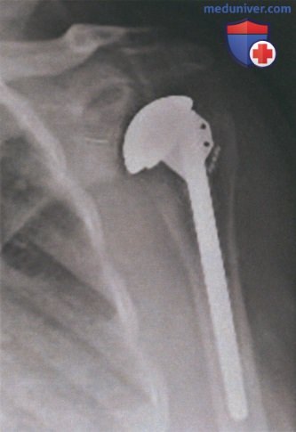
Рисунок 17
б) Список использованной литературы:
Braman JP, et al. The outcome of resection shoulder arthroplasty for recalcitrant shoulder infections. J Shoulder Elbow Surg 2006;15:549-53.
Cagle PJ, et al. Subscapularis repair after shoulder arthroplasty. Tech Shoulder Elbow Surg 2015;16(1):16-23.
Coste JS, et al. The management of infection in arthroplasty of the shoulder. J Bone Joint Surg [Br] 2004;86:65-9.
Debeer P, Plasschaert H, Stuyck J. Resection arthroplasty of the infected shoulder: a salvage procedure for the elderly patient. Acta Orthop Belg 2006;72:126-30.
Dilisio MF, et al. Arthroscopic tissue culture for the evaluation of periprosthetic shoulder infection. J Bone Joint Surg Am 2014;96(23):1952-8.
Flatow EL, Bigliani LU. Tips of the trade. Locating and protecting the axillary nerve in shoulder surgery: the tug test. Orthop Rev 1992;21:503-5.
Frangiamore SJ, et al. Neer Award 2015: analysis of cytokine profiles in the diagnosis of periprosthetic joint infections of the shoulder. J Shoulder Elbow Surg 2017;26(2):186-96.
Galatz LM, et al. Pectoralis major transfer for anterior-superior subluxation in massive rotator cuff insufficiency. J Shoulder Elbow Surg 2003;12:1-5.
Gavriilidis I, et al. Pectoralis major transfer for the treatment of irreparable anterosuperior rotator cuff tears. Int Orthop 2010;34:689-94.
Grosso MJ, et al. Sensitivity of frozen section histology for identifying Propionibacterium acnes infections in revision shoulder arthroplasty. J Bone Joint Surg Am 2014;96(6):442-7.
Klepps SJ, et al. Anatomic evaluation of the subcoracoid pectoralis major transfer in human cadavers. J Shoulder Elbow Surg 2001;10:453-9.
Matsen 3rd FA, et al. Origin of Propionibacterium in surgical wounds and evidence-based approach for culturing Propionibacterium from surgical sites. J Bone Joint Surg Am 2013;23:1811-7.
Nagda SH, et al. Neer Award 2005: peripheral nerve function during shoulder arthroplasty using intraoperative nerve monitoring. J Shoulder Elbow Surg 2007;1(3, Suppl. 1 ):6. S2-8.
Norris TR, Kelly JDI, Humphrey OS. Management of glenoid bone defects in revision shoulder arthroplasty: a new application of the reverse total shoulder prosthesis. Tech Shoulder Elbow Surg 2007;8:37-46.
Nwawka OK, Konin GP, Sneag DB, et al. Magnetic resonance imaging of shoulder arthroplasty. HSS J 2014;10:213-24.
O‘Driscoll SW, Petrie RS, Torchia ME. Arthroscopic removal of the glenoid component for failed total shoulder arthroplasty: a report of five cases. J Bone Joint Surg Am 2005;87:858-63.
Pottinger P, Butler-Wu S, Neradilek MB, et al. Prognostic factors for bacterial cultures positive for Propionibacterium acnes and other organisms in a large series of revision shoulder arthroplasties performed for stiffness, pain, or loosening. J Bone Joint Surg Am 2012;94(22):2075-83.
Rispoli DM, Sperling JW, Athwal GS, et al. Pain relief and functional results after resection arthroplasty of the shoulder. J Bone Joint Surg Br 2007;80:1184-7.
Scalise JJ, lannotti JP. Bone grafting severe glenoid defects in revision shoulder arthroplasty. Clin Orthop Relat Res 2008;466:139-45.
Shin JJ, et al. Pectoralis major transfer for treatment of irreparable subscapularis tear: a systematic review. Knee Surg Sports Traumatol Arthrosc 2016;24:1951-60.
Sperling JW, et al. Infection after shoulder arthroplasty. Clin Orthop Relat Res 2001 ;382:206-16.
Sperlinq JW, et al. Magnetic resonance imaging of painful shoulder arthroplasty. J Shoulder Elbow Surg 2002;11:315-21.
Stevens NM, Kim MH, Armstrong AD. Functional outcomes after shoulder resection: the patient's perspective. J Shoulder Elbow Surg 2015;24(9):247-54.
Venjakob AJ, et al. Arthroscopic removal of a polyethylene component in total shoulder arthroplasty. Arthrosc Tech 2015;4:49-52.
Weber P, et al. Management of the infected shoulder prosthesis: a retrospective analysis and review of literature. Int Orthop 2011 ;35:365-73.